Saturday, 11 August 2018

Class 12th Notes On Fertilisation & Double Fertilisation

                                            Fertilization 


In Angiosperm , fertilization or syngamy  is the process of interaction  of two opposite gamete for the formation of  the zygote  which  later develops into an embryo .   After fertilization the ovule forms the seed while ovary converts into fruit .

Process -  The  pollen grain  of the flower flow through the  air or attached to the   insects and reach  to the flower . This pollen grain  starts to germinate on the stigma of the female gametophyte and the pollen tube arises to develop through the style and reach to the ovule of the embryo sac . This pollen tube contains  two cells ; generative cells and vegetative cells . The vegetative cells have a reserved food material through which generative cell mature and divides.  Later on changed into  two male gamete cells .

 Now , the pollen tube enters into the  embryo sac through the micropyle region into one of the  synergid cells by  penetrating the filiform apparatus and deliver two male gametes while the other synergid cell will disintegrate  .  One male gamete combines with the egg to form zygote  , this later on   develops into the embryo similarly  the other male gamete fuses with the polar nuclei and form a  triploid cell or  Primary Endosperm Nucleus (PEN ) .

The union of two male gametes with the different nuclei of the embryo is known as Double Fertilization which is first discovered by the Russian scientist    S. G. Nawaschin in 1898  in Lilium and fritillaria .

Friday, 10 August 2018

7 Celled 8 Nucleate Structure Ovule With Labelled Diagram

                       Process Of Megasporogenesis In Angiosperm


In the previous blog, we had talked about how Archesporial cell meiotically divides and form four haploid cell arranged in a linear manner.
 Now out of the four cells, only one is functional while the other three degenerate. This is known as monosporic development.  One functional megaspore will give rise to the female gametophyte.
 Now let's go in more detail.

The functional megaspore will now start to divide mitotically and form two nuclei which start to move in the opposite direction where they form two nucleate embryo sac. Now the resulted two nuclei again divide by mitotic division and in this way, there is a formation of four nucleate and on further it divides more to becomes 8 nucleate of the embryo sac.

These 8 nucleate cells arranged in so well manner to form female gametophyte. Out of the eight nuclei, six nuclei covered by a cell wall and organized to form cells and the remaining two are known as Polar nuclei, situated below the egg apparatus in the central cell.

Now out of the six-cell, three cells organized in the embryo sac to form egg apparatus in the micropyle region of the ovule. This egg apparatus itself consists of two synergid cells and one egg cell. The synergid cell is consists of filiform apparatus, a finger-like projection help in inserting male gamete through a pollen tube.
At the Chalazal end, the three cells combine to form Antipodal cells, provide nutrition to the cell.
As I mentioned on the top that central cells have two polar nuclei, that's why after maturity this embryo sac has 8 nucleate, 7 celled structure. 


Thursday, 9 August 2018

The Structure Of Ovule : Biology Blog

                                    Megasporogenesis


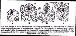
           This is the formation of megaspore through integuments ovule. Below the epidermis layer of the ovule, some cell continues to grow, out of this cell one cell enlarge known as Archesporial Cell or megaspore mother cell. This Archesporial cell meiotically divides to form four haploid megaspore cells, arranged in a linear fashion.

Before this I want to familiarize you with Ovule Structure, this makes a further understanding of the process easy for you.

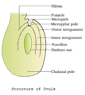
Ovule Structure - The typical ovule is originated from the placenta of the ovary which is small in structure and attached to the placenta by a stalk-like structure  Funicle. The area where the funicle and ovule fuses are known as Hilum.  On the outer side, two protective layers are present having a function to provide protection of the ovule, outer integument, and inner integument.  

There is one opening present at the region where inner integument arches over the nucellus known as the Micropyler region. On the opposite side we can see there is a fusion of outer integument and funiculus, this is the Chalazal End of the ovule.

The nucellus is the mass of the cell present inside inner integuments. The nucellus provides food material to the embryo sac which is present under nucellus.  

Wednesday, 8 August 2018

Class 12 notes on Structure of Pollen Grains and function

                                    Pollen Grains 


 What is Pollen Grain -

A very minute spherical structure present on the flower representing male gametophyte is known as Pollen Grains.  Pollen Grains are the medium for fertilization i.e., the tiny body is swirling in the air and attach to the female part and promote pollination. Pollen  Grains are approximately  25-50 micrometers in diameter.

Structure  - 

 Pollen has the outstanding feature of two-layered walls made in such a way that it shows a variety of patterns and at the same time also represents consistency in organization and chemistry.  The outer layer is known as Exine,  divided into two subparts, exine sexine, and exine nexin while  The inner layer is thin and made up of cellulose and pectin known as Intine. 

 Some chemicals are present on the outer wall of the pollen grains called  Sporopollenin which is one of the most resistant organic material and is thought to be responsible for the preservation of pollen in fossil plants deposit. The sporopollenin is absent in one place where some pores are present through this pollen tube emerge out during germination on stigma.

 The cell wall of the pollen grain is surrounded by a thin plasma membrane having distinct cytoplasm. After maturity, the cell of pollen contains two types of cell - vegetative and generative. The vegetative cell is having a  big size with an irregular nucleus. Their function is to provide food to the Generative cell and nourish them.

The Generative cell has a dense cytoplasm with a distinct nucleus. Coming to this stage some species of the Angiosperms  (60 to 70 percent)  ' pollen is shed off and this process is known as 2 -cell stage. The remaining species generative cells develop and divide mitotically and give rise to two male gamete cells and hence the 3 - cell stage.

Uses of Pollen -   

  • They are rich in nutrition supplements.
  •  They are also used in the form of medicine
  •   Store pollen can be used as a pollen bank just like a seed bank. 

We will cover the next part in our next blog, please subscribe to my blog for not missing any updates 

Top 10 biology book for class 12

                                      Top  10 Biology Books


Hello friends ,

 Are you looking for the best biology book to get success in  class 12th ?     
Friends , here I am suggesting you the top 10 biology book which you can buy from flipkart , with this I am also providing you direct link so its very easy for  you people to purchase .   


Tuesday, 7 August 2018

Life cycleof Angiosperm Overview : Biology Blog

                                        Angiosperms


What is Angiosperm -

Angiosperms ( flowering plants ) are the most advanced type of land plants having almost different sizes and shapes of a plant, it can be either small or it can be large also like Wolffia ( smaller ) and eucalyptus (large).  The ovule and the pollen grain are enclosed in a flower and the seed is enclosed in the fruit of these plants.
 If we talk about its usefulness, then it does not only provide food but also provides medicines,  fodder, fuel,  paper, and several other commercial products.

Life Cycle of Angiosperm -   The development of these plant is just like an animal that begins from a fertilized egg (sexual reproduction ) and converts into an embryo having a polarised organization, for example, its apical portion develop to from shoot, middle one into the stem and basal portion form root of the plant.   

L.S. Of Flower 
    Sexual reproduction consists of two ways - sporophytic ( microsporogenesis )  and gametophytic (megasporogenesis ). Sporophytes contain a diploid phase of a life cycle, the generation starts when the sperm nucleus fuse with the egg nucleus and another one fuses with the polar nuclei and form triploid endosperm and continue to form seed,  seedling, mature plants and flowers.  On the other hand, the gametophyte is having a very short haploid phase of the life cycle. Due to this the life cycle of Angiosperms is haplodiplontic type. 

Sporophytic (Microsporogenesis ) process - It is the process of formation of microspore with the help of pollen mother cells by the process of meiosis. This microspore further gives rise to the male gamete. 
The whole story of this begins with the flowering male reproductive part that is Stamen. This Stamen is generally consisting of two parts - Anther and the Filament. The size, color, and length of the anther can vary in different flowering plants.
The anther is generally having a bilobed structure having two thecae ( Dithecous  ). If we cut the anther transversely we found that the anther is having a tetragonal structure consisting of four microsporangia located in the corner of each lobe of the anther. After developing further this microsporangia give rise to pollen sac in which pollen grains are filled.

Mature Anther wall -   The typical microsporangium is looked like circular in the structure having four wall layers - the epidermis, endothecium, middle layer, and the tapetum. The outer three-layer protects the microsporangium and the last one, the innermost Tapetum nourishes the developing pollen grains. 
After some time some cells arrange in the center of microsporangium and form Sporogenous Tissue
.


We will cover the other half in our next blog post, please subscribe us to not miss any updates from biology blog

.

Monday, 6 August 2018

Structure & Function Of Secondary Oocyte

                                        Oocyte Structure


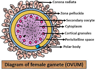
The egg or ovum is large in size as compared to the male sperm. It is non-motile and contains food in the form of yolk, glycogen, and protein. The ovum is composed of the following parts -

Plasmalemma

Primary membranes

Secondary membranes. 


  • Plasmalemma - It is the outer layer of the egg made up of lipoprotein. It is also known as the plasma membrane. This layer is 110 angstrom in thickness.

  • Primary membranes - In certain reptiles and coelenterate another egg membrane is also presently known as a primary or secondary membrane. It is secreted around the plasma membrane by the follicle cells of the ovary itself. In birds, molluscs, insects, this membrane is called by vitelline membrane while in mammals it is known as Zona pellucida. This vitelline membrane is made up of fibrous protein and mucoproteins. The plasma membrane and vitelline membrane are separated by some space known as Perivitelline space.

  • Secondary Membrane - The primary egg membrane is surrounded by another membrane for the protection of the embryo known as Secondary membrane. This membrane is secreted by oviduct and some reproductive organs of the body.  In amphibians, it gives jelly-like appearance while in reptiles, and in birds, the egg membrane and shell are secondary membrane. 

Related Links 

Types of Eggs | Amount Of Yolk | Distribution Of Yolk

                                             Types of Egg


Hello guys!

In this blog, we’ll explore a fascinating branch of zoology: the different types of eggs found in the animal kingdom. But before diving into the topic, let’s first understand what reproduction means.

Reproduction is a natural biological process through which parents produce offspring, typically of their own species. This process is essential for the survival and continuation of every species on Earth.

As we know, our planet is home to a vast range of species, each with its own unique way of producing eggs. Within these eggs lies the yolk — a rich source of nutrients. The yolk is made up of proteins, fats, vitamins, and minerals, all of which play a crucial role in the embryo’s growth and development.

Eggs are classified into different types based on the quantity of yolk they contain.

✨📚 Need Help with Notes or Assignments? 📚✨

Stuck with biology notes or struggling to finish your zoology assignment? Don’t worry — we’re here to help you learn better and score higher!

  • 💡 Personalized, topic-wise notes
  • 📝 Detailed assignment help
  • ⏳ Fast and reliable support

👉 Click here to get started!


l. On the basis of the amount of Yolk - Depending on the quantity of yolks, eggs can be 
  • Alecithal / Microlecithal / oligolecithal - The amount of yolk present in an organism's egg is of a very small amount or absent.   
      Example -  Sea urchin,  Amphioxus, Tunicates, Eutherian egg

  • Mesolecithal -  In this type of egg, the yolk amount is medium or moderate & concentrated towards one pole of the egg.
        Example -   Amphibian, Dipnoi fishes, lungfishes, Petromyzon

  • Polylecithal / macrolecithal / megalecithal -  The amount of yolk content is very large.         Example -    prototherian mammals,  insects, annelids, reptiles                
Types Of Eggs - Zoology


2. On the basis of the distribution of yolk - Depending on how they are distributed, eggs fall into one of the following categories:
  • Isolecithal / homolecithal Egg -   The yolk is equally distributed in the whole egg.Since the amount of yolk is relatively small, these eggs undergo holoblastic cleavage, resulting in the formation of cells of similar size during early embryonic development.
       Example -  Alecithal , micro, oligolecithal egg.

  • Teloleithal  Egg -  Here the yolk is found on the one pole of the egg. Usually, the egg has two opposite poles, one is the animal pole and the other one is the vegetal pole. Telolecithal types of the egg are those in which yolk is present on the vegetal pole while the other pole is free.
      Example -  amphibian egg.

  • Centrolecithal Egg -  

  • Centrolecithal eggs have a centrally located yolk, surrounded by a thin layer of cytoplasm and a large central nucleus. This type of egg is found in insects, such as flies and beetles. The yolk acts as a nutrient reserve for the developing embryo. Centrolecithal eggs undergo superficial cleavage, where cell division occurs only in the outermost layer of the cytoplasm, while the yolk remains intact.


tyoes of eggs

3. On the Basis of Shell - on the basis of a shell, there are two types of eggs -

  • Cleidoic Eggs -  The egg is surrounded by a hard covering or shell. These types of eggs are generally seen in terrestrial animals. 
          Example - reptile, bird
                                       
  • Non - Cleidoic Eggs -  The egg which is not surrounded by any hard covering.
           Example -  All viviparous animals.
                                                   
                                                 
4. On the basis of the development of egg - There are two different sorts of eggs based on how they develop:

Types Of Cleavage In Egg
  • Determinate /mosaic type - In some eggs, every part is already set with a specific role in the embryo's future development. If any part of the embryo is removed, the organism won’t develop properly. This is because each part is essential and can't be replaced or regenerated by the remaining sections. The missing portion cannot be recreated by the rest of the embryo. This kind of egg, where the fate of each part is already determined early on, is known as a determinate egg—it develops in a fixed, mosaic-like pattern.
          Example - annelids, molluscs.
  • Indeterminate type - An indeterminate egg is a type of egg found in certain animals, particularly reptiles and birds. It is characterized by having a high degree of developmental flexibility, meaning that the fate of the embryo is not predetermined at the time of fertilization. In indeterminate eggs, the blastomeres—the cells resulting from cleavage—do not have a fixed developmental fate, and each cell has the potential to develop into a complete and functional organism on its own.
          Example - amphibians.

Explore Top Blogs On Biology Topics

Aurelia Aurita Life Cycle: Characterstics, Classification Cell Cycle - Mitosis & Meiosis Difference between Prokaryotic & Eukaryotic cells | Biology Blog
Obelia Life Cycle, classification , Labelled  Diagram | Biology BlogKreb Cycle Steps With Diagram , Steps & ProductsPredator, Parasitic & Saprophytic Food Chain With Examples
What are the Important  features of SyconSycon Canal System With Diagram & Significance | Biology BlogDNA Transcription Process | Central Dogma | RNA Polymerase
 

Featured Post

Sycon Canal System With Diagram

                                     Canal system in Sycon  A sea creature called a sycon, or scypha , is usually found clinging to rock...