Friday, 3 August 2018

4 Phases Of Menstrual Cycle & Ovulation In Females

                                      Menstruation cycle


Every month, females undergo a natural reproductive cycle.

During this cycle, the body prepares itself for a possible pregnancy. If pregnancy does not occur, the body sheds the prepared uterine lining and resets the cycle for the next month. This process begins at puberty, a stage known as menarche, and continues until a woman reaches the age of approximately 40 to 50 years, when the cycle gradually comes to an end — a phase known as menopause.         
Age Of Menstruation


Duration - Generally, the duration of this cycle is ideally 28 days, but in some cases, its duration extends from 28 to 32.

                                         
Menstruation

Phases -  This cycle is completed in four phases -

  •       Menstrual Phase
  •        Pre-Ovulatory Phase
  •         Ovulatory Phase
  •          Post-Ovulatory Phase


  • 📘 Need Handwritten Notes or Custom Assignments on ovulation or Any Biology Topic?

    Struggling to understand tough biology topics like the menstrual cycle, types of eggs, or pollination? We offer clear, affordable, and ready-to-use handwritten notes and student-friendly assignments.

    📩 Mail Us for Notes
     Menstrual Phases -   During days 1-5, a low level of female sex hormone in the body causes the endometrium to disintegrate and its blood vessels to rupture. On day one of the cycle, a flow of blood and tissues, known as the menses, passes out of the vagina during menstruation, also called the menstrual period.

  • Pre-Ovulatory PhaseAfter about four to five days, the next phase begins, known as the follicular phase of the ovary. During this phase, the hypothalamus releases increasing amounts of GnRH (Gonadotropin-releasing hormone), which stimulates the anterior pituitary gland to secrete FSH (Follicle Stimulating Hormone). FSH promotes the growth and development of ovarian follicles, one of which matures into the Graafian follicle. As this follicle matures, the theca interna layer also begins to develop. The growing follicle starts secreting estrogen, a hormone that plays a key role in the proliferation (thickening) of the endometrial lining of the uterus, preparing it for potential implantation.

  • Ovulatory Phase -   The estrogen level continues to rise and reaches its peak by the end of the 10th day of the menstrual cycle. At this point, the high estrogen concentration sends a negative feedback signal to the anterior pituitary, causing it to reduce the secretion of Follicle-Stimulating Hormone (FSH). As a result, FSH levels begin to decline, while Luteinizing Hormone (LH) levels start to rise. This rapid increase in LH levels is called the LH surge. The LH surge triggers the rupture of the Graafian follicle, leading to the release of a mature ovum — a process known as ovulation, which typically occurs around the 14th day of the menstrual cycle.
  • Post Ovulatory Phase -   After the release of the ovum (secondary oocyte), the Graafian follicle undergoes degeneration and transforms into a new structure called the corpus luteum. This structure contains a yellowish fluid due to the presence of lutein pigment.

    If pregnancy does not occur, the corpus luteum begins to degenerate around the 14th day of the menstrual cycle. After degeneration, it is converted into a white scar-like structure called the corpus albicans, which contains a whitish substance.

    The formation and function of the corpus luteum are stimulated by the luteinizing hormone (LH). The corpus luteum secretes two important hormones: progesterone and estrogen. These hormones prepare the endometrium (inner lining of the uterus) for the implantation of the embryo by making it thick, vascular, and glandular.

    Progesterone plays a crucial role by inhibiting uterine contractions, thus helping maintain a suitable environment for pregnancy. If fertilization and implantation occur, the corpus luteum is maintained and continues to secrete progesterone.

    For this reason, progesterone is often called the "pregnancy hormone" because it supports the early stages of pregnancy.

                          
4 Phases of Menstruation Cycle


Menopause -Menopause is a natural stage in a woman's life when the ovarian and uterine cycles permanently stop. It typically occurs between the ages of 45 and 55. During this phase, the ovaries gradually stop producing the hormones estrogen and progesterone. At the onset of menopause, menstrual cycles may become irregular, but as long as menstruation continues, there is still a chance of conception. Therefore, a woman is considered to have officially reached menopause only after she has not had a menstrual period for 12 consecutive months.

Thursday, 2 August 2018

Explain Spermatogenesis & Mature Sperm Structure | Biology Blog

                                     Spermatogenesis


Spermatogenesis is the biological process through which sperm cells are formed from the germinal epithelium lining the seminiferous tubules of the testes. This epithelium consists of two main cell types:

  • Cuboidal primordial germ cells, which undergo division and differentiation to eventually become mature spermatozoa.

  • Tall somatic cells known as Sertoli cells (also called nurse cells), which support, nourish, and anchor the developing germ cells throughout the stages of spermatogenesis.

These Sertoli cells play a crucial role by creating a nurturing environment and regulating the development of sperm within the seminiferous tubules.

Spermatogenesis

📘 Need Handwritten Notes or Custom Assignments on Spermatogenesis or Any Biology Topic?

Struggling to understand complex biology topics like spermatogenesis, plastids, or transcription? We provide affordable, well-organized notes and biology assignments perfect for school and college students.

📩 Mail Us for Notes

Phases of spermatogenesis - It involves the following two phases -


A. Formation of spermatids - It includes the following phases -


  • 1. Multiplication Phase:
    During this phase, the sperm mother cells (also called spermatogonia) undergo mitotic division to produce new cells. Some of these cells migrate toward the lumen of the seminiferous tubules and enter the next stage—the growth phase—where they are referred to as primary spermatocytes.
    Other spermatogonia remain in their original position and continue dividing to maintain the pool of cells. These undifferentiated cells are known as stem cells.

    2. Growth Phase:
    In this phase, the primary spermatocytes grow in size. Their nuclei also enlarge, and they prepare for the upcoming maturation divisions.

    3. Maturation Phase:
    Each primary spermatocyte undergoes the first meiotic division (a reductional division), forming two haploid daughter cells called secondary spermatocytes.
    Each secondary spermatocyte then undergoes a second meiotic division (similar to mitosis), producing a total of four haploid cells known as spermatids.
    Thus, each primary spermatocyte ultimately gives rise to four spermatids, which will further differentiate into mature sperm cells.


B.Spermiogenesis – Transformation of Spermatids into Spermatozoa

The process of transforming spermatids into spermatozoa is known as spermiogenesis. The mature spermatozoa are commonly referred to as sperm.

Spermatids, which are produced after the maturation (meiotic) division, are haploid cells that resemble typical animal cells. However, in this form, they are non-functional as male gametes. Through the process of spermiogenesis (metamorphosis), these non-motile spermatids undergo a series of structural and physiological changes to become motile and functional sperm.

Key Changes During Spermiogenesis:

  1. Elongation of the Cell:
    The spermatid elongates lengthwise, increasing in size and acquiring the slender shape of a sperm.

  2. Nuclear Condensation and Shaping:
    The nucleus loses water from its nucleoplasm (nuclear sap), causing it to shrink and condense. It also takes on various shapes depending on the species.
    👉 The final shape of the nucleus determines the shape of the sperm head.

  3. Retention of the Cell Membrane:
    The original cell membrane of the spermatid remains intact and becomes the outer covering of the entire mature sperm, including the tail.

  4. Centriole Rearrangement and Tail Formation:
    The two centrioles in the spermatid rearrange themselves behind the nucleus:

    • The anterior centriole becomes the proximal centriole.

    • The posterior centriole becomes the distal centriole, which transforms into the basal body.

    • The basal body gives rise to the axial filament (or flagellum), which forms the core structure of the sperm tail, enabling motility.

Structure of motile sperm -

 The mature sperm consists of four parts -

  •    Head part 
  •    Neck
  •   Middle 
  •   Tail Part

                               
Sperm Diagram

  • The Head part consists of a dense nucleus in which cell organelles are present. On the tip of the nucleus, a small cap-like structure is presently known as Acrosome which is formed from the Golgi complex. It helps in penetration of Egg membrane during fertilization by dissolving enzymes called hyaluronidase.

  • The Neck part is a short segment of sperm that contains two centrioles, proximal and distal centriole. These are introduced at the time of fertilization along with the sperm nucleus to initiate cleavage in the zygote.

  • The middle part, Which is also known as the body of the sperm that contains mitochondria is present which function as to provide energy for the movement of sperm during copulation.
  • The last one is the Tail part which is the longest segment of sperm, consisting of central axial filament, the thin layer of cytoplasm, and an outer smooth plasma membrane. They have flagellar movement during copulation. 
Check - Male reproductive system in Hindi 

Difference between spermatogenesis and oogenesis
Difference between spermatogenesis & oogenesis

Hatch and Slack pathway or C4 cycle

                           Hatch and Slack Pathway / C4 Cycle

                                                 
 In 1967  Hatch and Slack proposed a pathway for dark reaction occur in sugarcane and maize plants. The first stable product is OAA, which is  4C, dicarboxylic acid. That is why the hatch and the slack pathway is also known as the C4 cycle. the detailed process is explained below -

                                       
Process -
  • The primary acceptor of carbon dioxide in C4 cycle plants is phosphoenol pyruvic acid (PEP). It accepts carbon dioxide and forms oxaloacetic acid (OAA).
  • OAA is the first stable product which is having four-carbon dicarboxylic acid. The reaction is catalyzed by PEP carboxylase. This is called beta carboxylation . This takes place in the mesophyll cell.
  • OAA is reduced to malic acid. The electron necessary for this reduction is supplied by NADPH. The enzyme malic dehydrogenase catalyzes the reduction.
  • The carboxylation of PEP and reduction of OAA takes place within the mesophyll cell.
  • Malic acid moves into bundle of sheath cells through plasmodesmata.
  • Within the bundle sheath cell Malic acid oxidatively decarboxylated into pyruvic acid.
  • The enzyme " malic enzyme "catalyze this decarboxylation. Carbon dioxide released in this reaction is accepted by RUBP. Carbon dioxide is fixed in the C3 cycle.
  • The electron released in the oxidative decarboxylation of Malic acid is accepted by NADP which is reduced to NADPH.
  • Pyruvic acid synthesized in the bundle of sheath cells moves into a mesophyll cell.
  • In the mesophyll cell, the pyruvic acid is phosphorylated to regenerate PEP. The enzyme pyruvic dikinase catalyzes the reaction. Two molecules of ATP are utilized in this reaction.  
Further Reading - Photosynthesis 
                                citric acid cycle 


Wednesday, 1 August 2018

Conducting system of the heart : ECG - BiologySolution

                                   Conducting System of the Heart 


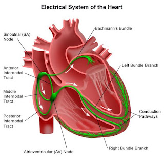
    The right and left bundle branches of the AV node, the SA node, the Purkinje fibre, and the AV bundle make up the heart's conductive system.


  • The sinoatrial node (SA) located in the wall of the right atrium near the superior vena cava.  The specialized muscle fiber that makes up this structure is unique that they can continually and mathematically send impulses ( send signals to contract )without any stimulation from the nervous system. This means the SA node is said to be self-exciting and that's the reason why the "SA node is known as the natural pacemaker of the heart."
  •  Now, before directly enters into the  AV node, the electrical impulse from the SA node is spread on to the right and left atria and gets depolarized. Due to this both the atria (right &left) contract simultaneously.
  • When the impulse reaches the AV node, there is a slight delay that allows the atria to finish their contraction before the ventricles begin their contraction.
  • The signal for the ventricles to contract passes the signal through the atrioventricular bundle. This bundle is differentiated into right and left bundle branches that conduct the impulse to the apex of the heart.
  • Therefore the signals are then passed to Purkinje fiber towards the ventricles to upwards myocardial.
  • The AV bundle, its branches, and the Purkinje fiber consist of specialized cardiac muscle fibers that efficiently cause the ventricles to contract. 
          In this way, the electrical conductivity of the heart can be recorded in the form of ECG.


ECG -  The electrical alterations that the myocardium experiences throughout the cardiac cycle are captured by an electrocardiogram (ECG). Ions that conduct electrical currents are present in our body fluids, therefore the myocardial electrical changes can be felt on the skin's surface. A device that monitors electrical changes in the heart is connected by wires to electrodes implanted on the skin during the ECG procedure.
Indirect proof of blood flow to the cardiac muscles can also be obtained via the ECG, which can also quantify the heartbeat's rhythm and pace.


  Working -           

  • After applying lead to the patient the machine started working. Now when the electrical signal is getting from the atria to ventricles it starts to depolarise. This is represented by  P.
  • P-Q segments show the time interval from traveling to SA node to the AV node.
  • QRS  merge and show ventricular depolarization.
  • Q wave shows the depolarization of the interventricular septum.
  • R wave produces by the main mass of the ventricle.
  • S phase shows the last phase of ventricular depolarization at the base of the heart.
  • Atrial repolarisation also occurs at this time.
  • S-T segment shows myocardial action potential. This is when the ventricle supplies the blood and pumps the heart.
  • T wave shows repolarisation immediately before relaxation. This cycle repeat with every heartbeat.


Understanding Bacterial Growth Curve: Phases | Biology Blog

                                    Bacterial Growth Curve 


In any biological system growth can be defined as the orderly increase of all chemical component . Increase of mass weight not really reflect growth curve because of the cells could be simply increasing their content of storage product such as glycogen . The idea of the bacterial growth curve was developed through careful observations and experiments done by early scientists in the late 1800s. They studied how bacteria grow and divided their growth process into different stages.Bacterial growth in batch culture can be modeled with four different phases -       
                      
  •       Lag Phase 

  •       Exponential Growth Phase

  •       Stationary Phase   

  •        Death Phase           
                                                                  

  •              Figure of Growth Curve
    Bacterial Growth Curve


  1. Lag Phase -     During Lag Phase , bacteria adapt themselves to growth condition . It is the period where the individual bacteria are maturing and not yet able to divide . During this period of bacterial growth cycle synthesis of RNA , Enzymes and other molecules occur .

  2. Exponential Growth Phase -  This phase is also known by another name called Log Phase . It is a period characterize by  cell doubling . The number of new bacteria appearing per unit time is proportional to the present population . If growth is not limited , doubling will continue at a constant rate , so both the number of cell and the rate of population increases double with each consecutive time period . For this type of exponential growth , plotting the natural logarithmic of   cell number against time produces a straight line .

  3. Stationary Phase -   The growth rate slow as a result of   nutrient depletion and accumulation of toxic product . This phase is reached as the bacteria begin to exhaust the resources that are available to them . This phase is a constant value as the rate of bacterial growth is equal to the rate of bacterial death .

  4. Death Phase -      Bacteria run out of nutrient and die . Death result from a number of factor ;  an important one is depletion of the cellular reserves of energy . like  growth , death  is a exponential function and hence in a logarithmic plot , the death phase is a linear decrease in number of viable cells with time . 
Also Read - Types of Eggs of Animals in Zoology (Embryology )

Tuesday, 31 July 2018

Brief notes on Oogenesis with 3 phases


                                                                     Oogenesis


The process of formation of the female egg is known as oogenesis. The process takes place in ovaries. Generally,  in most higher organisms,  this process starts when the female fetal is developing in the womb. The whole process can be studied in three phases -    

  • Multiplication Phase 
  • Growth Phase 
  • Maturation Phase            

OOgenesis
Read also -oocyte structure in human being 

  • Multiplication phase -  In the fetal embryo, the ovum mother cell or we can say oogonia start to multiply through mitosis and a  large number of diploid oogonia i.e., millions of oogonia are produced.


  • Growth Phase  Like spermatogenesis, oogonia grow in their size and shape to form primary oocyte with a diploid number of chromosomes. If we talk about other higher animals except for human then this phase is one of the largest phases of oogenesis. During the growth phase, several changes occur in egg and all these changes are classified into two subcategories -
         -  previtellogenesis
         -  vitellogenesis

  1. Previtellogenesis -   A number of changes occur in the nucleus and cytoplasm like the number of nucleoplasm increases, the nucleolus increases. Likewise, the number of cell organelle increases in cytoplasm, specially Golgi body, ER, mitochondria become increases in large numbers. Later all these three organelles arrange in a ring-like structure around the nucleus and there is a formation of the Balbiani Vitelline Ring.  Space appears in between the plasma membrane of the egg and vitelline membrane called perivitelline space in which a fluid is filled called perivitelline fluid.

  2. VitellogenesisNow egg starts storing food in the form of yolk. 


  • Maturation Phase - The primary oocyte starts its meiotic division in the fetal embryo but gets arrested in prophase the first stage of meiosis.   In this way, in a newborn girl, there is 2 lakh primary oocyte present in her ovary. When a girl attains a puberty stage than one of the ovaries stimulates only one primary oocyte for resuming meiosis first with the help of follicular stimulating hormone or FSH.  After completing meiosis first, the primary oocyte form two cell one is larger than another cell. Larger cell is known as a Secondary Oocyte and smaller one is known as Polar body. This secondary oocyte again started meiosis second but get arrested in metaphase second stage of meiosis. It then stops further and waits for the sperm to complete the further process of the meiotic division. After the entrance of sperm in the fallopian tube cell cycle again started by breaking the M phase promoting f tor or MPF and turning on the anaphase-promoting complex. Completion of meiosis second converts the secondary oocyte into a fertilized egg or zygote and also a second polar body.
What Is Ovulation?

Featured Post

Sycon Canal System With Diagram

                                     Canal system in Sycon  A sea creature called a sycon, or scypha , is usually found clinging to rock...