
Conducting system of the heart : ECG - BiologySolution

                                   Conducting System of the Heart 


    The right and left bundle branches of the AV node, the SA node, the Purkinje fibre, and the AV bundle make up the heart's conductive system.


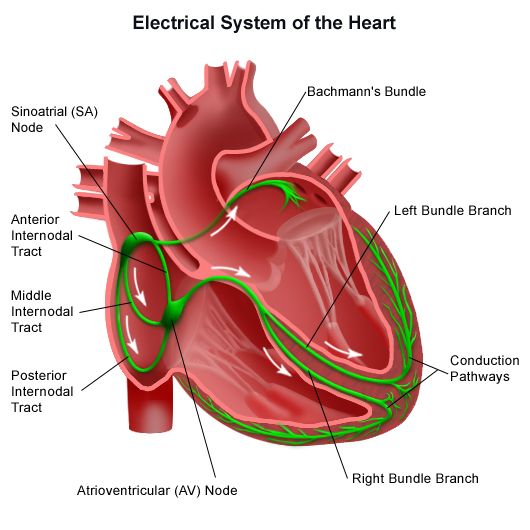
  • The sinoatrial node (SA) located in the wall of the right atrium near the superior vena cava.  The specialized muscle fiber that makes up this structure is unique that they can continually and mathematically send impulses ( send signals to contract )without any stimulation from the nervous system. This means the SA node is said to be self-exciting and that's the reason why the "SA node is known as the natural pacemaker of the heart."
  •  Now, before directly enters into the  AV node, the electrical impulse from the SA node is spread on to the right and left atria and gets depolarized. Due to this both the atria (right &left) contract simultaneously.
  • When the impulse reaches the AV node, there is a slight delay that allows the atria to finish their contraction before the ventricles begin their contraction.
  • The signal for the ventricles to contract passes the signal through the atrioventricular bundle. This bundle is differentiated into right and left bundle branches that conduct the impulse to the apex of the heart.
  • Therefore the signals are then passed to Purkinje fiber towards the ventricles to upwards myocardial.
  • The AV bundle, its branches, and the Purkinje fiber consist of specialized cardiac muscle fibers that efficiently cause the ventricles to contract. 
          In this way, the electrical conductivity of the heart can be recorded in the form of ECG.


ECG -  The electrical alterations that the myocardium experiences throughout the cardiac cycle are captured by an electrocardiogram (ECG). Ions that conduct electrical currents are present in our body fluids, therefore the myocardial electrical changes can be felt on the skin's surface. A device that monitors electrical changes in the heart is connected by wires to electrodes implanted on the skin during the ECG procedure.
Indirect proof of blood flow to the cardiac muscles can also be obtained via the ECG, which can also quantify the heartbeat's rhythm and pace.


  Working -           

  • After applying lead to the patient the machine started working. Now when the electrical signal is getting from the atria to ventricles it starts to depolarise. This is represented by  P.
  • P-Q segments show the time interval from traveling to SA node to the AV node.
  • QRS  merge and show ventricular depolarization.
  • Q wave shows the depolarization of the interventricular septum.
  • R wave produces by the main mass of the ventricle.
  • S phase shows the last phase of ventricular depolarization at the base of the heart.
  • Atrial repolarisation also occurs at this time.
  • S-T segment shows myocardial action potential. This is when the ventricle supplies the blood and pumps the heart.
  • T wave shows repolarisation immediately before relaxation. This cycle repeat with every heartbeat.


Comments

Popular posts from this blog

Sycon Canal System With Diagram

                                     Canal system in Sycon  A sea creature called a sycon, or scypha , is usually found clinging to rocks or other surfaces. These creatures have tube-shaped bodies that open outward with the help of a hole called an Osculum . They have tiny openings called Spiracles and Ostia all over their bodies to let water in. This water flows through canals inside their bodies and comes out through the Osculum, which is a larger opening used for breathing. We'll observe the shape of this canal system, why it is essential for those animals, type of canal system , and  its significance. The Sycon Type Canal system is the unique feature of the phylum Porifera . This system is also known as the  aquiferous canal system . This system supports the significant function of the body like nutrition, respiration, excretion, etc. History Overview -  Gran...

Whittaker's Five Kingdom Classification | Biology Blog

                                   Five Kingdom Classification Early biologists classified all species as either plants or animals. Since this method makes it difficult to research specific creatures, American taxonomist R.H. Whittaker created the five-kingdom classification in 1969. This system divides all organisms into groups based on how they are organized and how they are fed. Important standards for this classification include - The complexity of cell structure i.e. Prokaryotic and Eukaryotic. The complexity of the Organisms body is Unicellular and Multicellular. Mode of nutrition Autotrophs and Heterotrophs. Lifestyle. Phylogenetic Relationship. Based on these criteria, Whittaker classified the living world into the following categories  - Kingdom Monera                 Kingdom Protista Kingdom Fungi Kingdom Animalia Ki...

Aurelia Aurita Life Cycle: Characterstics, Classification

                                             Aurelia  Aurelia aurita is a type of zooplankton found all over the world. They are see-through and don't have eyes, brains, or hearts. They're usually between 2 to 15 inches wide. Even though they prefer being alone, they get carried together in groups called Blooms by wind and water currents. At night, they look like they're moving through the water like jellyfish, which is why they're sometimes called " jellyfish ".  They have four reproductive organs surrounded by tentacles in their top part. Their body color changes based on what they eat, so they can be blue, pink, or other colors. Let's learn more about Aurelia in detail. Details Of Aurelia Diagram -  Body : The center of the diagram would display the jellyfish's bell-shaped body, often called the " umbrella. " Tentacles : Below the bell, you'd see lon...