Posts

Showing posts from April 26, 2020

Explain principle of electron microscopy : Biology Blog

Image
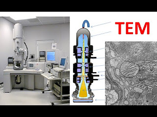
                              Principle of  electron microscopy Around 1931-32, two German scientists, Knoll and Ruska invented transmission electron microscopy and developed a transmission the electron microscope (TEM) around 1940. Principle - A strong beam of electrons discharged from tungsten filaments after supplying them with high voltage electronic current, pass through a vacuum and refocussed electromagnets. Radiation (wavelength 0.05 angstrom) is produced from it which is utilized as a source of illumination to the objects. An image is formed when electrons strike on a fluorescent screen or on a photographic film. Several hundred thousand times magnifications and resolution of about 5 angstroms can be achieved by TEM.  The compound of a transmission electron microscope - Electrogun - it is located on the top of the microscope. It works as a  source of electrons. It consists of tungsten filament surrounded by a negatively biased shield with an aperture from

Northern blot technique Of RNA - Principle , Steps | Biology Blog

Image
                                           Northern blotting   Hello guys! In this blog, we are going to explore the  northern blot technique . This technique can be used in any viral detection test as the genetic material of the virus is RNA. So, before moving to northern blot we focus on the word - blot. Blo t is basically a technique in molecular biology and genetics in which we DNA/RNA or proteins could be transferred onto the carrier membrane. The nylon membrane or nitrocellulose membrane are used for blotting purpose.  The transferring of molecules like DNA / RNA or proteins to the membrane can take place after gel electrophoresis. After blotting the transferred molecules are visualized.  For visualization, we can use the following methods-  1. If the probe is labeled with the radioactive chemical (32P), it can be observed on X-ray film directly.  2. If a chromogenic detection is used, it can be visualized by the development of color on the membrane its