Thursday, 24 July 2025

Understanding Population in Ecology: Attributes, Growth Models & Formulas

Understanding Population: A Complete Guide for Students

Population is one of the foundational topics in biology and geography, especially for students in middle and high school. It refers to the total number of individuals of a species living in a particular area at a given time. But population isn’t just about numbers—it also includes various characteristics, trends, and models that help us understand how species interact with their environment.

What is Population?

A population is a group of individuals of the same species that live in a defined geographical area and share or compete for similar resources such as food, water, shelter, and mates. These individuals also interbreed.

In biology, we study population ecology to understand the dynamics of species in their environment. In geography and demography, population studies help us plan cities, public services, and resources.

Population Attributes

Populations are described not only by their size but also by several attributes that influence their growth and structure:

  1. Population Size (N):
    The total number of individuals in a population.
    Example: N = 200 sparrows in a park.

  2. Population Density (D):
    The number of individuals per unit area.
    Formula:

    D=NAD = \frac{N}{A}

    Where:
    NN = Number of individuals,
    AA = Area

  3. Natality (Birth Rate):
    Number of births in a population over a specific time.

  4. Mortality (Death Rate):
    Number of deaths in a population over a specific time.

  5. Age Distribution:
    The proportion of individuals in different age groups:

    • Pre-reproductive

    • Reproductive

    • Post-reproductive


      Representation of age pyramid for human
  6. Sex Ratio:
    Ratio of males to females in the population.

  7. Growth Rate (r):
    The rate at which the population grows.

    📥 Get Detailed Handwritten Notes on Population (With Formulas & Diagrams)

    • Population formulas and solved examples
    • Graphs, population models, and attributes explained
    • Important theory points for quick revision
    • Class-ready explanations in simple, student-friendly language

    📧 Mail us now at bionotesandassignments@gmail.com to get the full PDF version – perfect for your exam prep!


      Formula:
  8. r=(BD)Nr = \frac{(B - D)}{N}

    Where:
    BB = births,
    DD = deaths

Population Growth Models

There are two major models to explain how populations grow:

1. Exponential Growth Model

  • Occurs when resources are unlimited.

  • Population increases rapidly.

  • Formula:

    Nt=N0×ertN_t = N_0 \times e^{rt}

    Where:
    N0N_0 = initial population,
    rr = growth rate,
    tt = time,
    ee = natural exponential base (~2.718)

2. Logistic Growth Model

  • Occurs when resources are limited.

  • Population grows rapidly at first but stabilizes at the carrying capacity (K).

  • Formula:

    dNdt=rN(KNK)\frac{dN}{dt} = rN \left( \frac{K - N}{K} \right)

    Where:
    KK = carrying capacity

Population Interactions

Populations don't live in isolation. They interact with other populations and their environment:

  • Competition – for food, space, etc.

  • Predation – predator-prey relationships.

  • Parasitism – one benefits, the other is harmed.

  • Commensalism – one benefits, the other is unaffected.

  • Mutualism – both species benefit.

Real-Life Applications of Population Studies

  • Urban Planning – for housing, transportation, and sanitation.

  • Wildlife Conservation – managing endangered species.

  • Healthcare Systems – understanding age and sex distribution for policy planning.

  • Agriculture – estimating pest populations to prevent crop loss.

Conclusion

Population studies help us understand how species grow, interact, and survive. Whether you’re learning biology or social science, understanding population attributes and growth models gives you tools to think about the environment scientifically and responsibly. Keep in mind the balance between nature and population for a sustainable future!


Tuesday, 22 July 2025

Understanding Mineral Nutrition in Plants: A Simple Guide for Students


Understanding Mineral Nutrition in Plants: A Simple Guide for Students

Mineral nutrition is a crucial aspect of plant biology, essential for growth, development, and reproduction. Every plant requires both macronutrients and micronutrients, absorbed from soil, water, or air, to perform basic physiological functions. In this guide, we’ll break down the methods used to study mineral requirements in plants, from ash analysis to hydroponic cultures, and understand the criteria for essential elements.

Understanding mineral nutrition in plants


Why Do Plants Need Minerals?

Plants, like all living organisms, need energy and nutrients for survival. While they produce carbohydrates via photosynthesis, minerals are absorbed from the soil and serve as building blocks for proteins, enzymes, nucleic acids, and other vital compounds.

Minerals are needed in specific quantities. Macronutrients like nitrogen (N), phosphorus (P), and potassium (K) are required in large amounts, whereas micronutrients such as zinc (Zn), boron (B), and manganese (Mn) are needed in smaller doses but are equally critical.

Methods to Study Mineral Requirements in Plants

1. Analysis of Plant Ash

One of the most fundamental techniques used to determine mineral content is plant ash analysis.

  • Fresh plant material is dried at 70–80°C to remove all water content.

  • The dried matter is then weighed to obtain its dry weight.

  • This dry matter contains polysaccharides, proteins, organic acids, and fats.

  • It is then burnt in a furnace at around 600°C, which removes all organic components by oxidation, releasing gases like CO₂, NH₃, and SO₂.

  • What remains is plant ash, composed solely of mineral elements.

  • The ash is then analyzed to determine the types and quantities of minerals present.

👉 However, this method does not reveal how the minerals are utilized or whether they are essential for plant survival.

2. Sand Culture Experiment

This is a more advanced technique to study the effect of individual mineral deficiencies:

  • Sterile sand is prepared by washing it with HCl, removing all natural minerals.

  • It is then washed with distilled water.

  • Plants are grown in this clean sand, and nutrient solutions are added.

There are two groups:

  • Control plants: Given all essential minerals.

  • Deficient plants: One or more nutrients are deliberately left out.

The difference in growth and deficiency symptoms helps determine the role of specific minerals.

👉 Common deficiencies observed include:

  • Yellowing leaves (chlorosis): Often due to nitrogen or magnesium deficiency.

  • Stunted growth: May be due to phosphorus deficiency.

3. Water Culture (Hydroponics)

Developed as early as 1860 by Sachs, this technique involves growing plants without soil, using a nutrient-rich water solution.

  • All required minerals are dissolved in water.

  • Roots are submerged in this solution.

  • It allows precise control over nutrient intake and is widely used in both labs and commercial agriculture.

Hydroponics helps study:

  • How individual minerals affect growth.

  • Interaction between multiple nutrients.

  • Alternative methods of plant cultivation in limited soil areas.

Criteria for Essentiality of an Element

Not all elements found in a plant are essential. There are specific criteria used to determine whether a mineral is essential:

  1. Absolutely necessary: The plant cannot complete its life cycle without it.

  2. Direct role in metabolism: Must be part of key biological processes, such as forming structural or functional molecules.

  3. Cannot be replaced: Its function is specific and cannot be substituted by another element.

  4. Lack causes visible symptoms: Deficiency must result in clear and correctable disorders.

  5. Reversibility: Supplying the missing element should correct the deficiency.

List of 17 Essential Elements

These elements are classified as either macronutrients or micronutrients:

  • Macronutrients: C, H, O, N, P, K, Ca, Mg, S

  • Micronutrients: B, Mn, Fe, Zn, Cu, Mo, Cl, Ni

Each of these plays a unique role in plant health. For example:

  • Nitrogen (N): Essential for amino acids and proteins.

  • Phosphorus (P): Important for energy transfer (ATP).

  • Potassium (K): Helps in enzyme activation and water regulation.

Modern Alternatives: Vermiculite

Sometimes, growing plants in sand can be challenging. That’s where vermiculite comes in—a mineral heated in a furnace to make it lightweight and porous.

Advantages of Vermiculite:

  • Sterile and chemically inert.

  • Higher water-holding capacity than sand or soil.

  • Does not degrade easily and can be reused.

Used widely in cutting plants and seed germination setups, vermiculite has become a reliable medium in experimental and commercial plant growth.

🌱 Conclusion

Studying mineral nutrition in plants helps us understand which elements are essential, how deficiencies affect growth, and what methods can best reveal these insights. From ash analysis to hydroponics, every technique offers a unique perspective. For students, grasping these methods builds a solid foundation in plant physiology, useful for exams, research, and beyond.

Explore Top Biology Blog Topics

Understanding Blood, Rh Factor & Lymph – Biology Guide What The Doctor Say About Cord Blood Banking
Mendelian Genetics : Biology Blog Function Of Essential Amino Acid
Chargaff's Rule of DNA | Biology Guide Obelia Life Cycle, Classification, Labelled Diagram
Types of Eggs | Amount Of Yolk | Distribution Of Yolk 4 Phases Of Menstrual Cycle & Ovulation In Females
Steps Of Glycolysis EMP Pathways – Biology Blog DNA Fingerprinting Procedure, Advantage & Application

Friday, 18 July 2025

Muscle Types, Structure & Proteins Explained Simply


All About Muscles: Types, Structure, and Contractile Proteins Explained

The act of movement, or changing position by one or more parts of the body, is a vital feature of living organisms. The study of movement is called Kinesiology, while the scientific study of muscles is known as Myology or Sarco-logy. Muscles are specialized tissues that help us perform different kinds of physical activity—from walking to heartbeat regulation.

myosin


Let’s understand the types of muscles, structure of muscle fibers, and contractile proteins that make movement possible.

Types of Muscles

Muscles can be classified into three main types:

  • Voluntary or Skeletal Muscles

  • Visceral or Smooth Muscles

  • Cardiac Muscles

Let’s begin with voluntary muscles, which we can control by our will.

💪 Need Detailed Notes on Muscle Types & Proteins?

Confused between skeletal, cardiac, and smooth muscles? Want clarity on actin, myosin, sarcomere structure and muscle contractions?

Get concise and exam-ready notes—perfectly explained for quick understanding!

👉 Mail us at: bionotesandassignments@gmail.com

Subject: Muscle Types & Structure Notes Request

  • 🧠 Simplified Diagrams & Tables
  • 🔍 Key Differences Highlighted
  • 📌 Suitable for: NEET • Boards • Assignments
  • 🕒 Fast Response | 💯 Trusted by Students

Voluntary Muscles

  1. Skeletal muscles constitute around 40 to 50% of the total body mass in an average healthy adult.

  2. They are connected to the skeletal system and are therefore also called skeletal muscles.

  3. These muscles show transverse lines (striations) at regular intervals under the microscope.

  4. As their contraction is controlled by our will, they are also called voluntary muscles. These muscles help animals (including humans) move, making them also known as locomotory muscles.

  5. Skeletal muscles are attached to bones by a tough cord of connective tissue called a tendon.

Structure of Muscle Fibre

Each muscle is made up of muscle fibers which are cylindrical or tubular in shape. The outer membrane of a muscle fiber is called the sarcolemma.

  • Inside the sarcolemma, each muscle fiber contains multinucleated cytoplasm, known as sarcoplasm.

  • The sarcoplasm is filled with myofibrils, which are fine thread-like structures arranged in parallel.

  • Each myofibril has alternate dark and light bands known as:

    • I-band (Isotropic Band) → contains actin filaments

    • A-band (Anisotropic Band) → contains myosin filaments

These bands give muscle fibers their characteristic striped (striated) appearance.

  • In the center of each I-band is a thin Z-line, which bisects the band and helps in the structural arrangement of the myofibrils.

  • The A-band contains thicker filaments and has a fibrous membrane at the center called the M-line.

  • The portion of the myofibril between two successive Z-lines is known as a sarcomere, which is the structural and functional unit of muscle contraction.

Sarcomere = 1 A-band + two half I-bands

Structure of Contractile Proteins

Two major proteins are responsible for muscle contraction: Actin and Myosin.

Actin Protein

  • Each actin filament is composed of two F (filamentous) actin chains that are helically wound around each other.

  • F-actin is a polymer of G (globular) actin units.

Tropomyosin

  • Tropomyosin is a type of contractile protein that lies in the groove of the actin helix.

  • In the relaxed state, it covers the active sites on actin, preventing unwanted contractions.

Troponin

  • Troponin is a complex of three subunits:

    • Troponin I

    • Troponin T

    • Troponin C

  • It is attached to one end of the tropomyosin molecule and is essential in the calcium-binding process that triggers muscle contraction.

Myosin Protein

  • Each myosin filament is also a polymerized protein made of several monomers.

  • A monomer of myosin (also called meromyosin) has two parts:

    1. A globular head with a short arm

    2. A tail called heavy meromyosin (HMM)

  • The globular head is enzymatically active. It contains ATPase activity and binding sites for both:

    • ATP (provides energy)

    • Actin (binds for contraction)

  • The short arm functions as a cross arm, helping the myosin head form a bridge with actin during contraction.

How Muscle Contraction Works

Muscle contraction is a result of the sliding filament theory, which states that actin filaments slide over myosin filaments, shortening the sarcomere and contracting the muscle. This process requires calcium ions (Ca²⁺) and ATP for energy.

During contraction:

  • Calcium binds to troponin C

  • This shifts the tropomyosin, exposing actin’s binding sites

  • Myosin heads attach to actin and pull the filaments inward

  • This results in muscle shortening and movement

Conclusion

Muscles are fascinating biological machines that help in movement, posture, and even vital functions like heartbeat and digestion. Understanding their types, structure, and the role of contractile proteins helps us appreciate how the body performs complex movements with precision.

From the skeletal muscles that allow us to walk and write, to the cardiac muscles that beat without rest, each muscle type plays a crucial role in survival. The study of muscle fibers and proteins like actin, myosin, tropomyosin, and troponin provides insight into how energy is converted into motion inside our bodies.

So the next time you stretch, walk, or run—remember the amazing biology that makes it possible!


🔗 Top Internal Links on Biology Blog

Topic Link
Types of Muscles: Structure, Proteins, and Functions Read Now
What is Pollination? Types and Mechanism Read Now
Short Notes on Anatomy of Cockroach Read Now
Why Are Living Organisms Classified? Read Now
7-Celled 8-Nucleate Structure Ovule With Labelled Diagram Read Now

Thursday, 17 July 2025

Understanding Blood, Rh Factor & Lymph – Biology Guide

Understanding Blood Components, Rh Factor, and Lymph: A Vital Look into Human Circulation

The human body is a complex system, and one of the most essential components sustaining life is blood. Blood not only transports oxygen and nutrients to various parts of the body but also plays a crucial role in defense, waste removal, and temperature regulation. Let’s explore the vital components of blood, the significance of Rh factor in transfusions and pregnancy, and the role of lymph in maintaining body balance.

Understanding blood , Rh factor & Lymph


What Is Blood?

Blood is a special connective tissue composed of a liquid matrix called plasma and formed elements such as red blood cells (RBCs), white blood cells (WBCs), and platelets.

Plasma:

Plasma is a straw-colored liquid that constitutes about 90–92% water and 6–8% proteins. These proteins include fibrinogen, globulins, and albumin.

  • Fibrinogen plays a key role in blood clotting.

  • Globulins are involved in the defense mechanism of the body.

  • Plasma also contains vital minerals (Na⁺, Ca²⁺, Mg²⁺, HCO₃⁻, Cl⁻), glucose, amino acids, lipids, and hormones.

Plasma without clotting factors is known as serum.

Formed Elements of Blood:

Erythrocytes (Red Blood Cells - RBCs):

RBCs are biconcave in shape and lack a nucleus. Their red color is due to hemoglobin, an iron-containing protein essential for oxygen transport.

  • An average healthy individual has about 12–16 gm of hemoglobin per 100 ml of blood.

  • The lifespan of RBCs is about 120 days.

  • They are produced in the bone marrow, and a healthy adult has about 5–5.5 million RBCs per cubic mm of blood.

Leukocytes (White Blood Cells - WBCs):

Also known as WBCs, these are colorless and fewer in number compared to RBCs.
They are broadly classified into:

  • Granulocytes (Neutrophils, Eosinophils, Basophils)

  • Agranulocytes (Lymphocytes, Monocytes)

  • Neutrophils are phagocytic in nature and help fight infections.

  • Eosinophils and Basophils play roles in allergic responses. Basophils secrete histamine, serotonin, and heparin, which aid in inflammation and immunity.

  • Lymphocytes (T-cells and B-cells) are critical to the immune system, helping in antibody production and cell-mediated immunity.

  • Monocytes are also phagocytic and help fight pathogens.

Platelets (Thrombocytes):

Platelets help in blood clotting.

  • A normal count ranges between 1,50,000–3,50,000 per mm³.

  • A decrease in platelets can lead to clotting disorders.

Rh Grouping and Its Importance

The Rh antigen was first discovered in Rhesus monkeys, hence the name. If this antigen is present on a person’s RBCs, they are termed Rh-positive (Rh⁺); if absent, they are Rh-negative (Rh⁻).

If an Rh⁻ person receives Rh⁺ blood, their body produces antibodies against the Rh antigen, which can cause complications. Therefore, blood group compatibility is crucial before transfusion.

Erythroblastosis Fetalis

Erythroblastosis fetalis is a serious condition that occurs when there is Rh incompatibility between a Rh⁻ mother and a Rh⁺ fetus.

  • During pregnancy or delivery, fetal Rh⁺ blood cells may enter the mother's bloodstream, leading her immune system to produce antibodies.

  • In subsequent pregnancies, these antibodies may cross the placenta and attack the fetal red blood cells, causing hemolytic anemia in the fetus.
    This condition is preventable with proper medical care and Rh immunoglobulin (RhIg) injections.

The Role of Lymph

Lymph is a clear fluid that enters the lymphatic capillaries from tissues. It is similar to blood plasma but has less protein content. It flows through lymphatic vessels and contains lymphocytes and granulocytes.

Functions of Lymph:

  1. Distributes nutrients and oxygen to tissue cells.

  2. Removes nitrogenous waste and CO₂.

  3. Destroys harmful pathogens via lymphocytes.

  4. Protects the body from diseases.

  5. Helps in maintaining body temperature.

Conclusion

The human circulatory system, comprising blood and lymph, ensures that every cell receives adequate nutrients and protection. Understanding components like plasma, RBCs, WBCs, and the Rh factor is vital for safe medical practices, especially blood transfusions and pregnancy management. Similarly, the lymphatic system plays an understated yet essential role in immunity and homeostasis. Awareness of conditions like erythroblastosis fetalis emphasizes the need for preventive care and the marvel of biological compatibility in human life.

Top B.Sc Topics In Biology 

Herdmania's Life: Anatomy, Behavior, Reproduction, and HabitatShort note on PhotosynthesisDifference between Prokaryotic & Eukaryotic cells | Biology Blog
Obelia Life Cycle, classification , Labelled  Diagram | Biology BlogKreb Cycle Steps With Diagram , Steps & ProductsTypes of Eggs of Animals in Zoology (Embryology ) | Biology Blog 
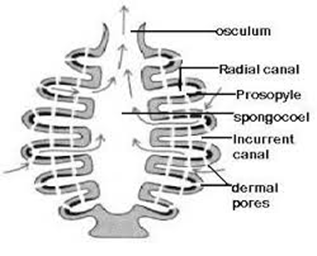
What are the Important  features of SyconSycon Canal System With Diagram & Significance | Biology BlogDNA Transcription Process | Central Dogma | RNA Polymerase


Friday, 11 July 2025

Plastids in Plant Cells: Types, Structure & Functions


Plastids in Plant Cells: Types, Structure & Functions

Introduction

Plastids are essential organelles found in plant cells and some algae. Coined by A.F.W. Schimper, the term "plastid" refers to various cell structures responsible for photosynthesis, pigment storage, and food storage. Understanding plastids—such as chloroplasts, chromoplasts, and leucoplasts—is fundamental in biology, especially for students preparing for exams in the UK and USA.

📚 Need Help with Biology Notes or Assignments?

Still confused about Plastids, Chloroplasts, or Cell Organelles? Looking for detailed notes or affordable assignments on biology topics?

👉 Mail us at bionotesandassignments@gmail.com to get ready-to-use biology notes at a student-friendly price!

What Are Plastids? | Definition & Importance

  • Plastids are large organelles found in all plant cells.

  • They are easily visible under a microscope.

  • Plastids contain pigments and contribute to essential plant processes like photosynthesis, color production, and storage of food.

Types of Plastids:

  1. Chloroplasts – for photosynthesis.

  2. Chromoplasts – store pigments responsible for red, orange, and yellow colors.

  3. Leucoplasts – colorless plastids for storage of starch, proteins, and fats.

Chloroplast: Structure, Function, and Importance in Photosynthesis

What Is a Chloroplast?

Chloroplasts are double-membrane, self-replicating organelles found in green plants. They contain chlorophyll pigments, DNA, and RNA, and they act as the plant's "photosynthetic kitchen."

Functions:

  • Main site for photosynthesis.

  • Synthesizes food using light energy.

  • Contains enzymes for carbohydrate and protein synthesis.

Structure:

  • Lens-shaped or oval.

  • Surrounded by a double membrane.

  • Contains thylakoids arranged in stacks called grana, suspended in a fluid called stroma.

  • Encloses a space called the lumen.

  • Ribosomes of 70S type present.

Chromoplast: The Pigment-Storing Plastids

Chromoplasts are responsible for the vibrant colors seen in fruits, flowers, and autumn leaves.

Key Features:

  • Found in light-exposed parts of the plant.

  • Contain carotenoids like carotene, xanthophyll, etc.

  • Store fat-soluble pigments giving red, orange, and yellow hues.

Leucoplast: The Storage Plastids in Plant Cells

Leucoplasts are colorless plastids that store nutrients in plant cells.

Types of Leucoplasts:

  • Amyloplasts – store starch.

  • Elaioplasts – store oils and fats.

  • Aleuroplasts – store proteins.

Additional Organelles: Peroxisomes and Glyoxysomes

Peroxisomes:

  • Contain enzymes like catalase.

  • Detoxify harmful substances like hydrogen peroxide.

Glyoxysomes:

  • Specialized peroxisomes found in fat-storing seeds (soybean, sunflower, peanut).

  • Convert fats into sugars during germination.

Spherosomes:

Tuesday, 8 July 2025

Explain What Is Pollination In Brief - Class 12th Bio Notes


   What is Pollination? Types, Mechanisms &                             Importance in Plants


What is Pollination


Pollination is one of the most essential processes in the life cycle of flowering plants. It refers to the transfer of pollen grains from the anther (male part) to the stigma (female part) of a flower, enabling fertilization and seed formation. This process ensures the continuation of plant species and promotes genetic diversity when it occurs between different plants.


🌼 Types of Pollination in Plants


Pollination in plants mainly happens in two ways:

• Self-Pollination

• Cross-Pollination


1. Self-Pollination

In self-pollination, pollen grains are transferred from the anther to the stigma of the same flower or another flower on the same plant. This type of pollination maintains genetic purity but reduces diversity. Self-pollination occurs in two main forms:

A) Autogamy

Autogamy happens when the pollen grains from the anther are transferred to the stigma of the same flower. It occurs through three methods:

• Cleistogamy: In some plants, the flowers do not open, ensuring complete self-pollination.

Examples: Oxalis, Viola.

• Homogamy: The anther and stigma of the bisexual flower mature at the same time.

Examples: Mirabilis, Potato, Sunflower.

• Bud Pollination: Anthers and stigma mature before the flower bud opens.

Examples: Wheat, Rice, Pea.

B) Geitonogamy

In geitonogamy, pollen grains are transferred from one flower to another flower on the same plant, but genetically it is still considered self-pollination.

🌸 Want Easy-to-Understand Pollination Notes?

Get high-quality, exam-ready notes on Pollination at a student-friendly price.

Still confused? Let’s make Biology simpler for you!

📩 Mail Us Now for Notes
Advantages of Self-Pollination

• Ensures higher chances of pollination.

• Maintains purity of the species and avoids unwanted mixing of traits.

• Plants do not need to produce a large number of pollen grains.

 Disadvantages of Self-Pollination


• No new desirable traits are introduced.

• Harmful characters cannot be easily removed.

• Does not support evolution and genetic improvement.

2. Cross-Pollination

Cross-pollination refers to the transfer of pollen grains from the flower of one plant to the stigma of another plant of the same species but genetically different individuals. It is also known as Xenogamy or Allogamy.

If you’d like to learn more about the structure of pollen grains, click here → Structure of Pollen Grains.

Natural Mechanisms That Promote Cross-Pollination:


a) Dichogamy

When the male and female parts mature at different times, preventing self-pollination.

Examples: Coriander, Jasmine, Sunflower.

b) Unisexuality

Male and female flowers occur on either the same plant (monoecious) or separate plants (dioecious).

c) Heterostyly

Flowers have different lengths of stamens and styles, preventing pollen from reaching the stigma of the same flower.

Examples: Primula, Linum.


d) Herkogamy

A physical barrier prevents self-pollination even when anther and stigma mature at the same time.

Examples: Calotropis, Orchids.

e) Self-Incompatibility

In some cases, pollen grains from the same flower fail to germinate on the stigma, preventing fertilization. This condition is called self-sterility.

🌸 Pollen-Pistil Interaction: The Journey of Pollen Grains

For pollination to result in fertilization, the pistil recognizes compatible pollen. Here’s how the process occurs:

1. Landing: Pollen grains land on a compatible stigma.

2. Germination: Pollen grains germinate, forming a pollen tube.

3. Pollen Tube Growth: The tube travels through the style toward the ovary.

4. Fertilization: Male gametes are released into the ovary, where fertilization takes place.


🌼 Artificial Cross-Pollination Techniques

Two important methods are used in plant breeding:

• Emasculation: Removal of stamens from a flower to prevent self-pollination.

• Bagging: Covering the flower with a bag to avoid unwanted pollination from other sources.


 Conclusion

Pollination is a crucial step in the life cycle of flowering plants. While self-pollination maintains genetic stability, cross-pollination brings genetic variation essential for evolution and adaptation. Understanding how plants control these processes helps in crop improvement and biodiversity conservation.


Monday, 16 December 2024

Explain Human Circulatory System in details

                     Human Circulatory System 


Human Circulatory system consist of heart , blood vessles & blood. Heart is mesodermal in nature. It is situated in the thoracic cavity , in between the two lungs. 
The double membrane that surround the heart is known as Pericardium. Pericardium circles the pericardial luid. Heart is 4 chamber with 2 upper small atria & 2 lower long ventricles. A thin wall seperates the left & right atria called Intra-atrial septum. The left & the right ventricles are seperated by thick Intra-ventricular septum

Human Circulatory System

The opening between the right atrium & right ventricles is guarded by a valve know as Tricuspid Valve. The Bicuspid Valve guards the opening between left atrium & left ventricles . The opening of right & left ventricles inton the pulmoery artry & the aorta. respectivelyare provided with the semilunalr valve. This valve in the heart allows the blood to flow in one dorection & thus preventing backflow of blood. 

Heart is a muscular organ. Haert muscle is known as Cardiac Muscle. A specialized cardiac musculature called the Nodal tissue is also dispersed in the heart. One is present in the upper left corner of the the right artrium called Av node

A bundle of nodal fibres, Av bundle contnues from the AVN which passes through the atria-ventricular septa & divides into right & left bundle. These branches give rise to minute fibres known as Purkinje Fibres. SA node is known as the Pacemaker of the heart as it has the ability to get excited & can genertaes an action potential.

Featured Post

Sycon Canal System With Diagram

                                     Canal system in Sycon  A sea creature called a sycon, or scypha , is usually found clinging to rock...